We are one of the largest Anthropology Departments in the UK and one of only a few to span Social Anthropology, Evolutionary Anthropology and Anthropology of Health. Our graduates have the opportunity to explore the full breadth of Anthropology or specialise in one of our sub-disciplinary areas. We are the only UK department to offer a residential field course (UK and international destinations) to all our undergraduate students.
QS World University Rankings by Subject 2024
We have been ranked 28th in the prestigious QS World University Rankings by Subject 2024.
The rankings, announced in March 2024 assessed over 19,100 subjects from almost 5,000 institutions across the world, based on academic and employer reputation, citations per academic paper, impact and quality of research and international research collaboration factors.
Durham is a world-leading university. Our academic excellence and impact is reflected in our global rankings and this is a true credit to the outstanding teaching and research carried out at our University.
Durham is consistently ranked as one of the world’s leading universities and among the top ten in the UK. It is ranked 78th in the QS World University Rankings 2024, 7th in the Guardian University Guide 2024 and Times and Sunday Times Good University Guide 2024.
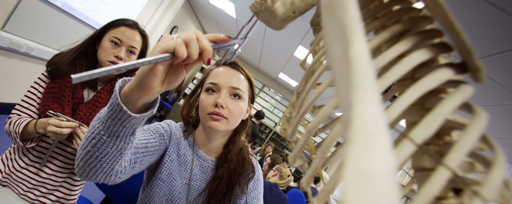
Our undergraduate courses introduce you to the comparative and evolutionary study of humans and draw on a range of social, cultural and biological approaches.
Our taught Masters offer you the opportunity to pursue advanced specialist courses and ‘conversion’ from other degrees. If you’re a PhD applicant, the sheer number of staff here means you are likely to find someone who can supervise and nurture your research interests.
Professor Jane Macnaughton from our Institute for Medical Humanities and Department of Anthropology has been chosen as the next Chair of the Nuffield Council on Bioethics Governing Board.
Scientists have long believed that big animals will tend to have big brains, but a new study involving Professor Robert Barton, from our Department of Anthropology, has found that may not be the case.
Innovator and Founder of Optim Energy, Jeremiah Thoronka has been featured in Forbes Africa’s 30 Under 30 list for his success and impact in the Climate Change and Sustainability (Social Impact) sector.
Hear from our students in the Department of Anthropology and follow us on social media.
Durham University
Contact us to find out more about undergraduate and postgraduate opportunities in our Department.
Durham UniversityDawson BuildingSouth RoadDurham, DH1 3LE
Check out our list of FAQs or submit an enquiry form.
Order your personalised prospectus and College guide here.