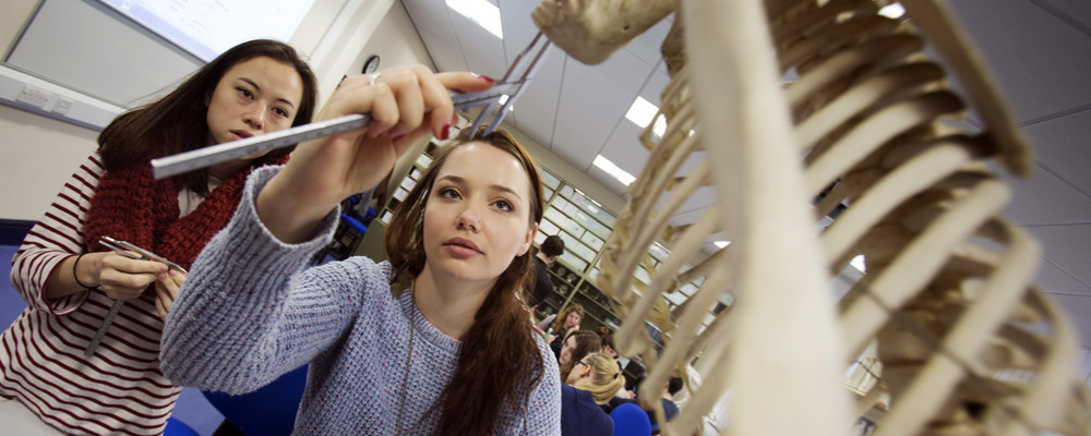
Durham Anthropology Journal
Volume 12(2-3) Copyright © 2005, Iwona Teul, Florian Czerwiski, Lech Cyryowski, Grzegorz Sawiski, Danuta Miklaszewska

Morphologic Evaluation of the Paranasal Sinuses of the Skulls from Different Historic Periods

Iwona Teul (1), Florian Czerwiski (1), Lech Cyryowski (2), Grzegorz Sawiski (1), Danuta Miklaszewska (1)

1. Departament of Anatomy, Pomeranian Medical University, 72 Powstaców Wielkopolskich str., 70-111 Szczecin, Poland

2. Departament of Diagnostic Imaging and Invasive Radiology, Pomeranian Medical University, 1 Unii Lubelskiej str., 71-252 Szczecin, Poland

teul@life.pl

Summary: The aim of this work was morphologic evaluation of the paranasal sinuses of the skulls from different historic periods. The study concerned skulls from early mediaeval period and from XIII-XVIII century. Evaluation of the size, shape and symmetry was performed on the basis of x-ray images performed in antero-posterior projection. Areas of the paranasal sinuses were calculated by means of appropriate software and correlations between chosen indices of the neurocranium and splanchnocranium were established.

In our material, differences in shape, symmetry, development level and presence of paranasal sinuses were stated. Our observations confirm that the frontal sinuses are the most changeable part of the pneumatic paranasal system and show great differences in shape, symmetry and degree of development. Bilateral absence of the frontal sinuses was observed mainly in early mediaeval skulls. In all studied skulls correlation between length, width and height of the neurocranium and height of the splanchnocranium with areas of the paranasal sinuses was observed. There is no correlation between maxillary sinuses area and height face index and upper face index and width of the maxilla.

Key words: paranasal sinuses, morphology, asymmetry, aplasia

1. Introduction

1.1. Pneumatisation of the skull is stricly connected with the paranasal sinuses: i.e., their morphology and degree of development. They constitute the first pneumatic system in human skulls; a second one is formed by the canals of the temporal bone and pneumatic mastoid process. It is an area which can show certain developmental directions of formation of the skull in human evolution. Anatomic, laryngologic, anthropologic, stomatologic and radiologic analysis can reveal diseases of our ancestors and significant changes in pneumatisation and symmetry are observed. The presence of the latter is an index of the stress: i.e., the reaction of the organism to life conditions according to modern anthropologic theories.

1.2. So far, no one in Poland has generally tried to estimate structure of the paranasal sinuses of people living in different climatic and enviromental conditions in different historic periods. Detailed analysis based on modern scientific methods enable evaluation of the skull pneumatisation in time and explain pathology of this part of the head. One has to remember that in times of great development of science and new technologies the osseous material is an inexhaustible source of knowledge about anatomic structure of a human being. Our study is supposed to tackle the problem of pneumatisation of the skull in time on the basis of material from different historical populations.

2. Materials and Methods

2.1. One hundred and three male early mediaeval skulls and 74 male skulls from XIII-XVIII century were included in our study. They originated from cemeteries in Cedynia (collection of Institute of Anthropology UAM in Pozna) and in Brze Kujawski (collection of Institute of Anthropology U in ód). All skulls had undamaged frontal and maxillary bones. There were no pathological or developmental changes found on any skull and their age was estimated as mature adults. Evaluation of the size, shape and symmetry of the sinuses was performed on the basis of x-ray images performed in antero-posterior projection. The line running through the superior margin of the orbit was taken as inferior border of the frontal sinus according to T. Koervelyessy's method while calculating of the area of the frontal sinuses. The areas were calculated by the means of computer software and statistically analysed (STATISTICA). Skull indices (width-length, frontal-width, width-length, face height, upper face, width of maxilla) were calculated according to Martin's metric code (Martin and Saller 1957). Then, correlations between chosen indices of the neurocranium, splanchnocranium and areas of the paranasal sinuses were established.

3. Results

3.1. In both groups of skulls, two kinds of the frontal sinuses were distinguished on the basis of shape: smooth and serrated. Additional division was based on presence or uni- or bilateral absence of the frontal sinuses. Fig. 1. Tab.I shows the division of the frontal sinuses according to shape, symmetry and degree of development. Presence and differences in size of left and right sinuses are shown in table II. On the base of data in table I and II results that in early mediaeval skulls from Cedynia and Brze Kujawski predominated the serrated frontal sinuses. Fig .2-3.

3.2. Uni- and bilateral aplasia of sinuses was observed more frequently in mediaeval skulls (Fig. 4). Arithmetic mean of the area of both sinuses in early mediaeval times was 6.018 cm2, and from Brze 7.884 cm2, which indicates greater area of sinuses in skulls from XIII-XVIII centuries. The length and width indices showed the highest correlation value. Calculated areas of maxillary sinuses in both groups reveal differences in their size. They are not so much different in shape as the frontal sinuses. Average area of the maxillary sinuses is 6.895 and 7.130 for Cedynia and Brze, respectively. No statistically significant differences between areas of left and right sinuses were observed. The maxillary sinuses do not show great variation in shape and formation. Correlations between their pneumatisation and width of the body of the maxilla, length of the splanchnocranium and length of the skull were established. Correlation with other indexes were less distinguished.

3.3. Evaluation of sinuses on the base x-rays does not give such accuracy as analysis of CT images, which gives more topographic details, especially concerning its internal structure. That is why the next step of our investigation will be comparing of x-ray and CT images in evaluation of paranasal sinuses.

4. Discussion

4.1. Importance of the paranasal sinuses is still controversial. The most popular theory is that their presence lowers the weigth of the skull and mechanically fixes bones of the splanchnocranium. Shock absorption / insulation/ and other protection for brain and other neural organs have also been posited. Other concept suggests lowering the pressure during mastication, warming, and control of pressure of inspired atmospheric air, amplifying of the voice power or smelling properties (Mooney and Siegel 2002). Comparing of radiological images of paranasal sinuses and clinical symptoms reveals connections that cannot be accidental. Among people complaining on chronic headache, there is a great percentage with excessive pneumatisation of the frontal sinuses. In case of some tumors lower the degree of pneumatisation of the maxillary sinus observed.

4.2. Anatomy of the frontal and maxillary sinuses was subject of many studies, some of them concerned their symmetry (Anagnostopoulou and Venieratos 1991), hypolasia and aplasia (Bolger 1990), dependence on the palatine cleft, malocclusions, development in different stages of life (Koppe and Yamamoto 1994) and influence of climate (Hanson 1980, Koertvelyessy 1972) on pneumatisation (Strk 1992).

4.3. Pneumatization proceeds independently of other cranial changes in early life. Some believe the sinuses play a role in development that goes beyond a mere peculiarity of ossification. Because maxillary sinuses formation is a result of the pneumatization of several bones, some researchers have suggested that its presence may be the result of variable allometric development. Supporting this latter view are the observations by Koppe and colleagues (1996) that the size of the maxillary sinus is related more closely to skull or body size than to more functionally related measures such as palatal width and lengh. Regardless, the sinus appers to develop rather independently early in life, but then becomes more coordinated in its growth with the maxilla (Koppe 1994, Mooney 2002).

4.4. Many feel that the morphology and function of early paranasal sinuses evolved as an adaptation to respiratory and olfactory needs and modification to the cranial vault. As the need for olfaction diminished during later primate evolution and the molars enlarged, changing the shape of the dental arches and other cranial elements, the sinuses came to be more involved physiologically with respiration and thermoregulation (Takahashi 1983).

4.5. It is supposed that the time played certain role in process of development of mastication organ and influence the size of paranasal sinuses. Additionally the studies of bone part from one cemetery can enrich general knowledge about less known population.

4.6. Absence of asymmetry of the maxillary sinuses is confirmed by other authors (Szilvassy 1981, Bolger 1990). In neither group was uni- or bilateral lack of the maxillary sinus observed. According to Bloger (1990), aplasia of the maxillary sinuses is stated in 0.5%. Lang and Hass (1988) observed hypoplasia of the frontal sinuses in 1.4%.(3). Mineo (1987) stated that aplasia of the frontal sinus occurs when their area doesn't exceed 0.8 cm2. Using this definition, bilateral lack of sinuses was shown in different male populations: among 25% Eskimoes from Alaska (Hanson 1980), 11% bushmen in Australia, and 13% of Japaneese (Koertvelyessy 1972). Strk and others (1992) studying skulls from XVI-XVI centuries observed aplasia of the frontal sinuses in 3.2%. In our study, uni- or bilateral aplasia of the frontal sinuses was observed. In their work, Jonas and Mann (1976) deny the correlation between asymmetry of the face and asymmetry of the paranasal sinuses. Our results differs from those obtained by Otkay (1992) for Turkish population. It is probably caused by the influence of the climatic conditions (Hanson 1980) and method of measurements he used. Aplasia of the paranasal sinuses is connected with smaller sizes of the skull.

5. Conclusions:

5.1. The frontal sinuses are the most changeable part of the paranasal apparatus contrary to the maxillary sinuses.

5.2. The early mediaeval skulls are less pneumatised than XIII-XVIII century skulls.

5.3. Pneumatisation of the left side predominates in both studied groups.

5.4. Bilateral aplasia of the frontal sinuses was more frequent in mediaeval skulls.

5.5. The length, height and width of the skull influence the size of the frontal sinuses.

5.6. The length of the skull, height of the splanchnocranium, width of the body of the maxilla influence the size of the maxillary sinuses.

References

Anagnostopoulou S., Venieratos D. (1991) Classification of human maxillar sinuses according to ther geometric features. Anat. Anz. 173: 121-30

Bolger W.E., Woodruff WW. (1990) Maxillarysinus hypoplasia:classification and description of associated uncinate process hypoplasia. Otolaryngol-Head-Neck Surg. 103 (5Pt1): 759-65

Davis WE., Temler J.,Parsons DS. (1996) Anatomy of the paranasal sinuses Otolaryngol Clin North Am 29: 57-74

Hanson Ch.L., Owsley D.W. (1980) Frontal sinus size in Eskimo populations. Am. J. Phys. Anthropol . 53: 251-255

Jonas I., Mann W., (1976) Die Kieferhohlenasymetrie als Ursache der Roengenologischen. Fehldiagnose einer Sinustis Lryng-Rhino-Otol 55:905-13

Koertvelyessy T. (1972) Relationships between the frontal sinus and climatic conditions: a skeletal approach to cold adaptation Am. J. Phys. Anthropol. 37: 161-172

Koppe T., Yamamoto T., O., and Nagai H. (1994) Investigationsnon the growth pattern of the maxillary sinus in Japanese human fetuses. Okajimas Folia Anat.Jpn. 71: 311-318

Lang J., Haas A., (1988) Über die Sagittalausdehnung des Sinus frontalis, dessen Wanddicke, Abstände zur Lamina cribrosa, die Tiefe der sogenanntenn Olfactorius-Rinne und die Canales ethmoidales Gegenbaurs. Morphol. Jahrb. 134: 459-469

Martin R., Saller K.( 1957) Lehrbuch der Antropologie. Gustav Fischer Verlag: Stuttgart

Mineo Y., Sachio M., Hajjme S. (1987) Classification system of frontal sinus patterns by radiography. Its application to inentification of unknown skeletal remains. Forensic. Sci. Int. 34: 289-299

Mooney M.P., Siegel M.I. (2002) Understanding craniofacial anomalies. John Wiley & Sons: New Jersey

Oktay H.(1992) The study of the maxillary sinus aresa in different orthodontic malocclusion Am-J-Orthod-Dentofacial-Orthop. 102: 143-145

Strk P., Kaczanowski K., Skawina A. et al. (1992) The morphological evaluation of frontal sinuses in the human skulls Folia Morphol. 51: 319-328

Szilvassy J. (1974) Die Stirnhöhlen der Schädel aus dem awarischen Gräberfeld von Zwölffaxing. Niderösterreich Ann Naturhist. Mus. Wien, 78: 109-118

Szilvassy J. (1981) Zur Entwicklung der Strinhöhlen. Anthropol. Anz 39,2: 137-149